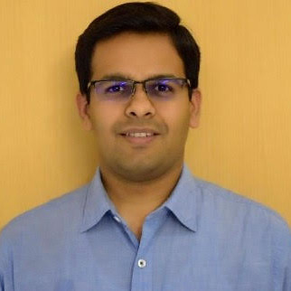
Faculty Name: Dr. Gunjan Mehta
Course Period: 31st July to 16th October 2023 (Monday 4:00PM to 5:30PM; Thursday 2:30PM to 4:00PM)
Number of Credits: 2 credits
Course Contents: Introduction to fluorescence microscopy and its applications; design of a fluorescence microscope and its optics; various illumination strategies such as wide field, confocal, TIRF/HILO, light sheet; fluorescent proteins, dyes, and fluorescence labeling strategies; 3D imaging; live cell imaging; time-lapse imaging; super-resolution microscopy (SIM, STED, STORM/PALM); single-molecule imaging; optical tweezers and traction force microscopy; high content imaging; techniques such as FRET, FRAP, FLIM, immunofluorescence, Fluorescence In-situ Hybridization (FISH), Spatial mapping of gene expression (RNAscope); digital images and camera technologies; introduction to Fiji/ImageJ, application of artificial intelligence/machine learning and virtual reality in image analysis.
The practical component of this course includes image processing, data analysis, quantification, and visualization using Fiji/ImageJ. Quantification of biological information from the microscopic images, intensity measurement, image segmentation, colocalization, quantification, and visualization of 3D images, deconvolution, 3D rendering, and reconstruction.
What you'll learn: Students will learn basics of fluorescence microscopy and its applications in life science/biology research. Different illumination strategies such as wide-field, confocal, light-sheet, TIRF, two-photon and various techniques such as FRET, FRAP, FLIM, immunofluorescence, 5D imaging, time-lapse imaging, single-molecule and super-resolution imaging etc. will be taught. Students will also learn basics of digital images and camera technologies used in fluorescence microscopy and how to quantify biological information from the digital images using Fiji/ImageJ.
About the Instructor: Dr. Gunjan Mehta is an assistant professor in the Department of Biotechnology at IIT Hyderabad. He used advanced imaging technologies to study cell division during his Ph.D. (2009-2015) at IIT Bombay. He worked as a Post-Doctoral Fellow (2015-2020) at the National Cancer Institute (NCI), National Institutes of Health (NIH), Bethesda, MD, USA. During his post-doc, he developed single-molecule imaging technique for quantifying the dynamics of proteins in live cells at the NCI optical microscopy core facility. He has more than 13 years of experience developing and applying fluorescence microscopy techniques for biomedical research. Currently, his research group at IIT Hyderabad studies the mechanism of chromosome dynamics and gene regulation using single-molecule imaging approaches.

None
There will be two exams (MCQs + short answer questions) of 25 points each, image processing assignment (25 points), and student presentations (25 points). Total 100 points.
Fee: Rs.20,000/- Plus GST
Note: The payment link will be shared only with the shortlisted candidates by email
